1The patient must be part of the explanation#
A tumour can shrink while the person living with it grows weaker. A treatment can reach its target while the patient gains little useful function. A person can meet the criteria for recovery and still lack the capacity to withstand the next illness.
For that person, success means being able to think, move, work, care for someone else, and make plans again. Medicine needs to explain the distance between a biological change and that return to life.
Patient-state medicine organises diagnosis, prevention and treatment around a person’s changing capacity to respond, recover and retain function. It asks what is failing in this patient, which intervention their physiology can use, and what must be restored for benefit to last.
Cirsentra Labs is a computational physiology lab building the scientific foundation for this approach. Our work connects original analyses of human biological data with experimental and clinical literature. From that work we are building a mechanistic physiological model of patient health: a connected account of how a patient’s state is produced, how it sustains disease, and how a treatment changes the function available afterwards.1 Its purpose is practical: to show which operation has to change, how to change it, and what the patient will be able to do afterwards.
Our ambition is to make differences between patients explainable, measurable and actionable. A diagnosis should lead to a treatment decision that accounts for the person who must live with its consequences.
2Give patient differences a physiological explanation#
Two people can share a diagnosis and need different interventions. The same inherited risk can lead to different disease trajectories. A treatment that helped last year can fail this year. Illness, treatment and incomplete recovery can change what a patient brings to a diagnosis that has not changed. An apparently modest illness can cause lasting decline in one person while another recovers.
Calling these differences patient variability names the problem. Explaining them requires following what happens between a cause and its effect.
A signal or drug must reach the right place. The receptor must be able to use it and pass it on. The tissue must have the energy and materials to carry out the response. Other organs must support that work. The response must end, its products must be cleared, and the capacity it consumed must be rebuilt.
Failure at each step creates a different problem and a different opportunity to intervene.
The patient’s state determines which paths from cause to consequence are available at that moment. A useful account of state therefore includes receiving capacity, energy, organ coordination, compensation and recovery. It also includes the history that made those capacities what they are.
This is the meaning of reserve geometry: the arrangement of capacities and constraints that determines how a cell, organ or person responds at a given time, what that effort costs, and what remains possible afterwards. One organ may maintain its output because another is doing extra work. A reassuring measurement can coexist with a growing cost elsewhere.
Within that arrangement, the disease substrate is the part currently producing or sustaining the problem. A detected receptor, an age-related molecular mark or an old exposure can belong to a patient’s state without being what now has to be corrected. Each process takes its role from the patient and the moment: one starts a disturbance, another keeps it going, another amplifies it, another compensates for it. One process can do two jobs at once. Extra insulin can hold blood sugar steady while changing what other tissues are exposed to.
The limit can also move. While a damaging exposure continues, it limits treatment. Once it is controlled, the depleted receptors become the limit. Once they return, the work the tissue has to do may be what remains. Treatment changes the state on which the next decision depends, so the plan can change while the diagnosis stays the same.
Patient-state medicine makes that cost, and the capacity behind the measurement, part of the explanation.
3Every response helps set the next one#
We set this out as the Reserve-Setting Law:
Each biological response and its recovery set the capacity available for the next demand.
The body that encounters the next infection, treatment or physical demand has been changed by what came before. It may have rebuilt what it used, gained capacity through adaptation, or carried a deficit forward.
Our work on biological state memory describes how such changes are kept. A tissue can appear healed yet hold a changed state that shows only when it is challenged again. That state can protect the next response, lock a problem in place, or change which cells respond next time.
Section 9 sets out how the law is tested in a patient: an equivalent demand, repeated after a defined recovery interval, with output, cost and recovery measured each time.
The reserve-setting cycle follows that change: what reaches the tissue, how it is received, what work is commanded and done, which systems compensate, how the response ends and is cleared, and what is rebuilt for the next demand.
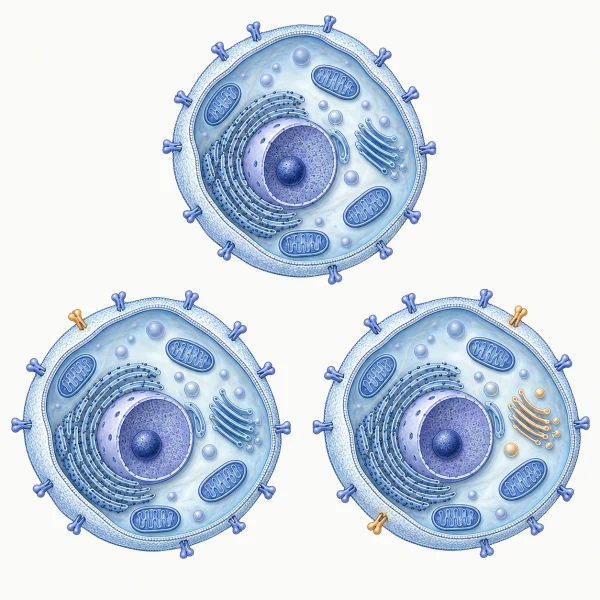
This gives recurrence and loss of treatment response a physiological question: what was available the first time that is no longer available now? It also gives prevention a task: identify the capacity being lost while the patient can still produce an apparently normal result.
Recovery becomes part of how the next outcome is produced. Treatment schedules, rehabilitation and repeat dosing have to account for what the previous response left behind.
Our work on the first reserve takes this sequence back to development before birth. The body constructs the vessels, receiving cells and systems for growth, defence and repair that later demands will encounter. The placenta does not simply pass on what is in the mother’s blood: it selects, converts, retains and clears what reaches the growing child. Construction continues after birth, as feeding, breathing, infection and repair first act on organs that are still maturing. Inherited biology and developmental conditions contribute to that starting architecture, and life continues to reshape it. Patients begin adult life at different baselines.
A patient’s future depends partly on what their body has already had to do, and how well it was able to rebuild.
4A treatment needs a receiver that works#
Much of medicine acts by sending an instruction: a hormone, a drug, a nutrient, a stimulus. Its effect depends on the receptor that receives it and the signalling that carries it onward.
We call that requirement the competent receiver. A receptor must be correctly built, accessible, intact, correctly placed and connected to the processes that carry its signal onward. Its continued availability and renewal decide whether the same response can be produced again. A receptor population can give a normal first response and a weak second one, because too little of it has returned to work before the next demand arrives.
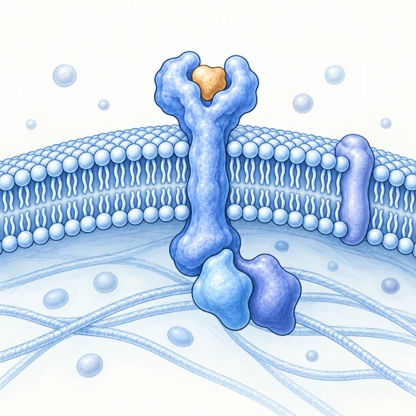
Counting receptors or measuring their gene expression answers part of this question. The physiological or pharmacological effect of an input depends on the currently usable receiver population and its renewal dynamics, not on gene expression or total receptor abundance alone. A target can be detectable while too little usable capacity remains to produce the required response.
Sepsis shows this in people. Natural killer cells stay present and countable in the blood while they cannot mount the response the infection calls for: their interleukin-12 receptor expression and their interferon-gamma output remain suppressed for at least two weeks after diagnosis. Blocking AMP kinase, the cell’s energy switch, restores that output in cells taken from those patients.2 The cells were there the whole time. What they lacked was the capacity to act, and the rescue names where it was lost.
This changes the intervention question. More signal helps when supply is low. When the receiver is impaired, treatment has to address that impairment or use another functioning route.
Reception and execution have different requirements. A tissue can receive an instruction correctly and still lack the energy to carry it out. Picard and Murugan named the property behind that limit: energy resistance, the resistance to electron flow through which a cell turns the potential held in food-derived electrons into useful work. Life needs that resistance, and too much of it produces oxidative stress, heat, inflammation and molecular damage. Their relation fixes the arithmetic: at a given energy potential, a tissue running at half its usual electron flux meets four times the resistance.3
Energy-constraint research describes the same pressure across the whole body: a finite budget shared among staying alive, handling stress, and growth, maintenance and repair.4 We take these principles into the clinic. When the work outruns the energy that funds it, other systems cover the gap, and the covering costs energy of its own. Measured output holds while that cost accumulates, so function can decline quietly and then fail quickly once demand crosses what the tissue can fund. Energy shortfall also slows the rebuilding of receivers, so the two losses feed each other, and energy can recover before function does. The failure has two seats that can be measured: the energy sensor inside the cell, and the blood-facing brain interfaces that read the distress signal a failing tissue exports, GDF15, and answer with a body-wide command to eat less, rest and move resources. That command is correct, and it still takes material away from rebuilding. The receptors can be intact at first, and the patient still deteriorates.
Mitochondria connect this energy account to the rest of the patient, and they do more than supply energy. In dividing cells, respiration is also needed to make aspartate, a building block for proteins and nucleotides, so a preserved energy supply does not mean a preserved capacity to build.5 Mitochondrial chemistry makes hormones: variants in FDXR, a mitochondrial enzyme that feeds electrons into steroid synthesis, cause adrenal insufficiency, and steroid-producing cells made from people with these variants produce too little of the adrenal steroid hormones.6 Stressed mitochondria change what a cell sends out. Human cells with defective energy machinery spend more energy and secrete more GDF15,7 and after injury, mitochondrial DNA and peptides released from damaged tissue activate immune cells.8 Mitochondria also have to be rebuilt, and a cell can regain its resting respiration before it can rebuild what later work needs.
Repairing the mitochondrial source, blocking the signal it sends and rebuilding the organelle are therefore different treatments, with different results to measure. Treatment aimed at a named maintenance process can already help: in an open-label study of 16 patients with TK2 deficiency, a disorder of mitochondrial DNA maintenance, function stabilised or improved on deoxynucleoside therapy, judged against historical comparisons.9
The clinical task is to tell these states apart. A shortage of signal, a damaged receiver and an energy-limited response call for different solutions.
5The body has an architecture for setting state#
The brain responds continually to the condition of the body, coordinating circulation, fluid balance, endocrine activity, immune responses, daily rhythms and behaviour. That coordination depends on specialised interfaces through which information passes between blood, brain and cerebrospinal fluid.
The anatomy is specific. The blood-brain barrier is intact across more than 99 per cent of the brain. A small set of structures are the constitutive exceptions: the area postrema, the median eminence, the subfornical organ and the vascular organ of the lamina terminalis, the choroid plexus, the pineal, and the neurohypophysis. Their vessels are fenestrated by design, so circulating signals meet neural tissue directly. Local cellular barriers regulate onward access and help define what each interface receives and transmits.10
Eight signals from the body reach the brain substantially through these interfaces. Together they report what the body has to work with and what it is dealing with: leptin and insulin for fuel and fat stores, insulin-like growth factor for growth and repair, thyroid hormone for the pace of metabolism, corticosteroids such as cortisol for stress, and interleukin-1, tumour necrosis factor and interleukin-17 for inflammation.
Single blood-vessel cells in the adult human hypothalamus carry the machinery for many of these signals at once. In 1,534 such cells from 11 donors, individual cells carried transcripts for up to nine signal routes, the eight above plus sex steroids, in graded combinations; 597 cells carried four or more. An independent cohort of 20,120 cortical vessel cells from 30 donors reproduced the pattern. We call these Convergence Cells. Cells that carry more routes also tend to carry more of the machinery that sets vessel tone, moves material and remodels the vessel. That machinery is counted as transcripts, not measured at work. Whether paired signals act on these cells together, and how they are arranged at the median eminence, are the next experiments.
Medicine already treats at these sites without naming them as a system. Antiemetics act at the area postrema. Fezolinetant acts at the arcuate nucleus for menopausal vasomotor symptoms. The appetite and nausea effects of the GLP-1 drugs arise at the same two sites. Their status as one coordinated reading surface is new.
One trial has tested this route directly. In cancer cachexia, stressed tissue secretes GDF15 into the blood, and the area postrema reads it through the GFRAL receptor. An antibody against GDF15 was given to 187 patients with raised GDF15. Every dose improved body weight at 12 weeks, by 1.22, 1.92 and 2.81 kilograms above placebo, and the highest dose also improved appetite and physical activity.11 Blocking one circulating signal changed what patients could do. The trial measured the signal and the patient; the link to the interface comes from anatomy. Where the GDF15 comes from, and whether the patient’s tissues rebuild once it is blocked, need their own measurements.
Together these sites and the interfaces related to them make one system, which we call the circumventricular sentinel network. Sentinel describes the work they do. These cells stand at the border, sample what arrives in blood and cerebrospinal fluid, set what the rest of the body does about it, and then read the result when the changed state returns to them. The network turns what it reads into hormones, nerve traffic, barrier changes, pressure and fluid balance, the timing of the day, and behaviour. Because a single interface controls several of those outputs, a change there is felt across several systems at once.
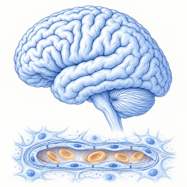
The same loop explains how disease can be sustained from the centre. A disturbance in the body changes what reaches an interface; the interface changes hormonal or nervous output; the body’s response changes what returns. Along that loop, an interface can modify how a signal acts, amplify the disturbance it causes, or keep it going after the original cause has changed. We call the proposed interaction at the vessel wall the Convergence Gate: one signal changes how the interface handles the next. Each of these roles is a hypothesis with a defined experiment: measure what arrives, what the interface does with it, what the body does next, and what happens when the returning signal is interrupted.
This is central to our account of patient state. It gives specific places to investigate how a problem elsewhere in the body changes brain function, and how a change in brain-body communication reshapes the condition of the whole patient. It also gives treatment new places to act: neutralise a harmful incoming signal, correct how the interface handles it, protect its receptors, or interrupt a reinforcing return.
6What reaches the system changes what it can do#
A receiving cell meets a changing environment: hormones, nutrients, medicines, inflammatory signals and microbial products. We call these external effectors because they act on the receiving system from outside it. Their sources lie inside the body and outside it.
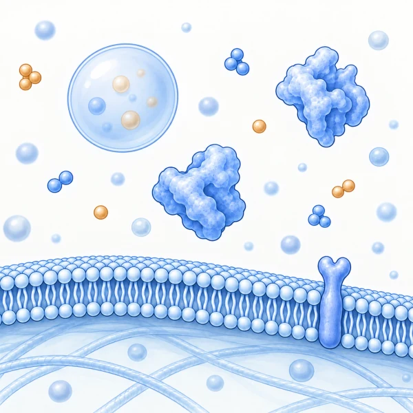
They reach tissue by several routes, and some of them cut and degrade the receptors that treatment depends on. We study one source of them closely: the bacteria of the mouth. Porphyromonas gingivalis, a keystone organism of periodontal disease, releases proteases called gingipains that digest host proteins for food. Published work shows these enzymes cut the insulin receptor, and that cells exposed to them take up less sugar.12 Whether membrane vesicles carry them to receptors elsewhere in the body is one of the questions we are testing.
Across organs, shared signals must produce different but compatible actions. One bloodstream carries the commands and the materials at once, and the liver, muscle, kidney and brain each owe that same instruction a different answer. We call this systemic metabolic readability: how an organ turns what reaches it from that one bloodstream into its own correctly timed answer, through arrival, receiver capacity, execution, compensation and recovery.
The two ideas answer different questions. Energy resistance asks whether one tissue can fund the work it was told to do. Readability asks whether the organs’ separate answers still fit together, and what it costs to hold them in line. Organs do more than compete for a shared energy budget.4 What one organ does changes what another receives. When muscle handles sugar poorly, the pancreas may release more insulin to hold blood sugar steady, and the liver and fat tissue then meet a different exposure. Their answer changes the blood again, and the materials used today build the receptors the next meal will need.
Health depends on those different responses working together. A hormone level alone cannot describe that coordination. It also takes knowing which tissues can use the signal, which must compensate, and how the work affects subsequent recovery.
Our findings on sex- and life-stage receiver architecture add another dimension: the receptors and signalling available for these responses differ between people and change across a life. Those differences belong in the explanation of disease and treatment response, and in the design of the measurements used to assess them. Sex can enter this architecture at several steps: through the dose of sex-chromosome genes, through hormone exposure during a developmental window, through the placenta, and through the mosaic of cells produced by X-chromosome inactivation. Each calls for a different measurement.
Together, these discoveries connect what reaches the patient, what the patient can do with it, and how the response changes what comes next.
7Why this layer has been difficult to see#
Modern biology resolves molecules, maps individual cells and reveals changes in gene activity at extraordinary scale. Clinical medicine measures organ function, disease severity and physiological reserve, and bedside judgement and performance scores record what the person can still do. Each provides a view of the patient, and connecting those views across organs and through time is a further scientific task.
Specialisation divides that task. A receptor is studied in one field, its circulating influences in another, the patient’s symptoms in a third, and recovery in a fourth. A measurement of abundance is available where a measurement of usable capacity is needed. A resting result is recorded where the decisive information is how the system responds and rebuilds.
Anatomy creates a further difficulty, and it is the deeper reason. A structure can occupy a unique causal position without expressing unique molecules. Topology can be the source of biological function. Any method that searches for molecular distinctiveness passes over a structure whose importance lies in where it sits, what reaches it, and where its output goes. The interfaces above are exactly of that kind, which is why they have been studied largely one at a time rather than as one coordinated system.
The same gap runs through computational biology. The State model from Arc Institute, the largest perturbation model built so far, was trained on nearly 170 million observed cells and more than 100 million perturbed cells across 70 cell lines.13 Every perturbation in that training was delivered outside a body. A cell in culture carries no endocrine axis, no immune context, no circulating protease load, and no interface that sets its state from the blood. A model trained this way predicts what a cell does when perturbed. It cannot say which patient is in a state where that perturbation helps. The layer is missing from the data because it had not been mapped as a system.
Clinical records have a version of the same gap. A diagnosis code and a visit date record no demand, no timing and no recovery, so a model trained on them inherits what the record left out. A model of patient state needs data that keeps them: when the demand arrived, what it cost, how long function took to return, and which source recorded it. It also has to keep the patient separate from the estimate. A new laboratory result changes what a model believes without changing the patient; a treatment can change the patient before the record shows it. A physiological model of patient health earns its name when its predictions are tested against what an intervention actually changes.
We use an AI-assisted knowledge graph to bring human data, mechanisms, disease trajectories, drug effects and source evidence into a queryable body of knowledge. AI retrieves and connects material at scale. Human scientific judgement directs the questions, weighs explanations and chooses the next analyses and experiments. This makes it possible to carry knowledge across disease boundaries while keeping the mechanism and the evidence in view.
8Connect the hallmarks to the whole patient#
The hallmarks of aging and cancer organise major changes in cells and tissues. We connect those changes to the capacities they alter, the work they transfer to other organs, and the state they leave behind.
Aging
Our hallmarks of aging work describes a reconstruction-attrition architecture: across repeated demands, the body rebuilds progressively less of the capacity it needs for the next response. Molecular damage, impaired clearance, receiver loss and energy constraints enter that sequence at different points. Other systems compensate, keeping a routine result stable while recovery takes longer and the range of tasks the person can manage contracts.14
An aging tissue carries its history, but it lives in conditions that can change. Heart transplantation separates the age of an organ from the age of the body supporting it: in mice, grafts shifted in molecular age measures toward their recipients, and a small study of human heart biopsies points the same way.15 The question for each patient is which present conditions preserve function, which sustain a restriction, and which can be changed.
This gives aging research a concrete objective: preserve the capacity to recover and respond again across time. An intervention should be assessed by the function it restores, the cost of maintaining it, and whether that capacity remains available at the next demand. The trajectory of recovered capacity becomes a treatment outcome.
Cancer
Our hallmarks of cancer work places the tumour in a changing host. A cancer’s capabilities depend on the signals and materials that reach its cells, the machinery that turns them into work, and its ability to maintain that machinery through repeated demands. The same holds for the host tissues that must contain it, tolerate treatment and recover between cycles. One treatment can suppress the tumour, preserve useful host function and create a new disturbance at the same time. In mouse tumour models, blocking the growth enzyme PI3K raised the host’s insulin, and that insulin restored the tumour’s growth signalling despite continued drug exposure.1617
This is why tumour response and patient recovery have to be followed together. A smaller tumour can coexist with a depleted host, and the next treatment meets both changed states. Treatment design has to ask which side has lost capacity, which side can rebuild, and what the next cycle will favour. The decisive comparison holds the early effect on the tumour comparable, changes one host operation, and measures later tumour control and host function separately.
9What should change for patients and clinicians#
A patient should be able to ask:
- What is preventing me from improving?
- What does this treatment require my body to be able to do?
- What needs to recover before the next treatment or demand?
- How will we know that I am regaining capacity for everyday life?
Patient-state medicine turns these questions into a diagnostic and treatment agenda. It calls for assays that distinguish failed delivery, impaired reception, limited execution and incomplete recovery; for treatment choices linked to those findings; and for follow-up that tracks whether useful capacity returns.
One test shows whether capacity returned. Measure the step that limits this patient under a defined demand, apply the repair that matches it, then, where it is safe, repeat an equivalent demand after a defined recovery interval. Compare output, cost and recovery each time. The first response shows what the treatment did. The second shows how much usable capacity the patient got back. A second response held only at a higher cost is a narrower reserve, not a restored one. A treatment that improves the first response and leaves the second unchanged has borrowed capacity rather than restored it.
Three results should be kept apart. Restored function means the task can again be done at an acceptable cost and recovery. Restored state means the biology behind it has also returned. Independence from support is a third result, tested only when withdrawing support is safe. Continuing support that keeps a person functioning is a valuable outcome in its own right.
For clinicians, the aim is a physiological reason for a decision: why this intervention, for this patient, at this time. Dose and timing become questions about drug exposure and about the tissue’s ability to respond. A poor response prompts investigation of the failed step and of the conditions needed for a better one.
A diagnostic test and a treatment have different jobs. A test must tell competing explanations apart; a treatment must improve what the patient can do. The order of treatment follows what currently limits the patient. While a damaging process still destroys receptors faster than they are replaced, making more of them leaves the population depleted: control the damage first, protect the receptors that remain, then rebuild the ones that were lost.
The patient’s account often holds the sequence that needs explaining. “I can get through the day, but then I need two days to recover” describes a demand, a response and a prolonged recovery. Recorded as fatigue, the account loses that sequence. Changes in endurance, clarity of thought, sleep and recovery show what to investigate. Measurements then test the explanation and follow whether treatment changes the problem the person lives with. Follow-up keeps a symptom recorded at one visit connected to the demands and recovery around it.
Performance status scales and the World Health Organization’s classification of functioning describe how limited a person is.1819 The next question is which constraint produced that limit, whether it can be changed, and whether changing it would let the person benefit from treatment. A reversible limitation becomes a treatment objective. Preparing a patient to respond, and leaving room to rebuild between demands, belong in the design of treatment.
Patient-state medicine also changes what trials and care pathways count as success. In oncology, tumour control and the patient’s ability to tolerate treatment and recover belong in the same account. In muscle loss, preserving tissue has to translate into useful strength and function. In brain disease and rehabilitation, the capacity to sustain recovery belongs alongside the immediate response.
The standard we seek is improvement that leaves the patient more able to live, and more able to meet what comes next.
10What should change for drug development#
Target-capacity pharmacology preserves, rebuilds, bypasses or diversifies usable target capacity, separately from changing ligand supply, occupancy, delivery or downstream pathway tone.
Its central sequence is:
- Drug exposure
- target engagement
- usable target capacity
- physiological response
- clinical benefit.
Each transition has requirements. Measuring whether a drug reaches and binds a target leads to the next question: does enough usable target capacity remain, in the right tissue and state, to produce the intended benefit?
That question changes what we develop. Alongside altering a signal or blocking a pathway, we can seek interventions that protect or restore the capacity a treatment needs. When the signal is excessive and the receiver still works, reducing the signal treats the cause. When the receiver itself is damaged, reducing the signal leaves that damage in place. It changes how we select patients: a trial can measure the state the drug’s mechanism requires, then test whether that state predicts benefit. It changes how we investigate failure: insufficient exposure, impaired target function and a downstream execution limit become distinct explanations to examine.
Medicine already has a precedent. In cystic fibrosis caused by the Phe508del variant, lumacaftor helps the faulty CFTR channel be processed and reach the cell surface, and ivacaftor makes the channel work. Together they improved lung function and reduced exacerbations in randomised trials.20 Building more usable target and changing its activity were complementary treatments. We ask the same question of every target a treatment depends on, and of whether it is still available at the next demand.
This makes restoring treatment responsiveness a development goal in its own right. When a therapy needs machinery the patient has lost, an enabling treatment can restore that prerequisite first. A trial can compare the therapy, the enabling treatment, both together and both in sequence, measuring the prerequisite just before the therapy is given. A drug that raises the first response while wearing down its target can leave the next response weaker, so screening follows the target through time, not only its first engagement.
This is not a theoretical distinction. In September 2026, a trial of pelacarsen in 8,323 patients with raised lipoprotein(a) and established cardiovascular disease lowered the particle the drug was designed to lower and did not meet its primary endpoint of major adverse cardiovascular events.21 A trial can lower the input it targeted and still miss its endpoint, because lowering the input was never the same as completing the repair. The question this framework puts to that result is whether reducing the particle changed the vascular state that produces events: whether clearance, repair and the thrombotic balance of the vessel wall recovered, and in which patients they still could. Full results have not been published.
The patient’s state also changes during treatment. Drug development therefore has to ask whether the response can be repeated, what recovery it requires, and whether improving one system imposes an unsustainable cost on another.
The opportunity is to make physiological fit a design criterion from target selection through clinical development. The same science guides new drug targets, companion assays, treatment combinations and intervention timing.
11What we are building#
Cirsentra began with a question that followed more than a decade of my work connecting basic sciences and clinical medicine across a full medical curriculum: how do we turn knowledge of separate mechanisms into an explanation of the person in front of us?
For the past two and a half years I have built and financed the research needed to pursue that question across diseases. The resulting laws, receiver concepts and maps of systemic coordination now join in one mechanistic physiological model of patient health, the common foundation for the next stage.
We are setting out to:
- Make patient state measurable. Develop assays that locate the failing operation and track the capacity to respond and recover.
- Make treatment fit explainable. Connect each intervention to the physiological conditions it requires, and test those conditions in patients.
- Make lost capacity a treatment target. Develop ways to preserve, rebuild, bypass or diversify the receivers and cellular machinery, including mitochondria, on which useful responses depend.
- Make central feedback treatable. Test the brain interfaces as places where disease is modified, amplified or maintained, and develop interventions that act there.
- Make lasting function an outcome. Follow recovery, repeated demand and everyday capability alongside disease-specific endpoints.
- Make the science cumulative. Publish the laws, supporting analyses and results of clinical tests so that others can examine, use and extend them.
The distance between how long people live and how long they function is often treated as a fact of nature. The World Health Organization put that distance at 9.5 years worldwide and 12.5 years in the United States in 2021.22 Closing it starts with naming which operation has failed in which patient.
This work needs clinicians, experimental scientists, assay developers, computational researchers and drug developers working across the same causal sequence. We invite them to help turn these discoveries into measurements and interventions that change care.
The future we are working towards is one in which a patient’s differences guide what happens next: what to treat, what to restore, when to act, and how to recognise recovery that lasts.
A person comes to medicine with a life they want to keep living. Patient-state medicine exists to make their capacity for that life part of every treatment decision.
Authorship and sources#
Patient-state medicine is the category through which Cirsentra Labs brings together its research on patient physiology, disease trajectories and treatment response. This manifesto states the scientific account and the clinical direction of that work.
The foundations described here draw on original computational analyses of human biological data and on published experimental and clinical research. Eleven foundational preprints are in preparation: reserve geometry, the reserve-setting law, biological state memory, prenatal reserve construction, the competent receiver, systemic metabolic readability, measuring deployable reserve, systemic signals at the brain’s vascular interface, energy resistance, external effectors and the executable disease substrate. An integrating paper, Patient-State Medicine: Building a Physiological Model of Patient Health, sets out how they connect, and two further preprints apply the framework to aging and cancer.[14][17] This document presents the connected argument for a wider readership. The papers provide the analyses and evidence for individual claims, and citations will be added here as each receives a permanent identifier.
1 The integrating paper. Seiler F. Patient-State Medicine: Building a Physiological Model of Patient Health. Physiological foundations for disease mechanisms, diagnosis, treatment, and recovery. Version 6.1, 29 September 2026. Preprint in preparation. It connects the eleven foundational preprints and sets out the experiments that test them.
2 Deployable immune capacity in sepsis. van der Wurff A, et al. Disturbed metabolic adaptation drives natural killer cell dysfunction in association with nosocomial infection during human sepsis. EBioMedicine. 2026;129:106345. doi:10.1016/j.ebiom.2026.106345. Interleukin-12 receptor expression and interferon-gamma output stayed suppressed for at least 14 days after sepsis diagnosis, and inhibiting AMP kinase restored mTORC1 activity and interferon-gamma production in cells from those patients.
3 Energy resistance. Picard M, Murugan NJ. The energy resistance principle. Cell Metabolism. 2025;37(11):2107-2127. doi:10.1016/j.cmet.2025.09.002. The principle and its relation are theirs. Reading it at the blood-facing interfaces, as a rate law for patient state with thresholds that can be measured in people, is Cirsentra’s work.
4 Energy constraint. Energy constraint on human health. Trends in Endocrinology & Metabolism. Published online 22 April 2026. doi:10.1016/j.tem.2026.02.010. The allocation of energy among vital, stress, and growth, maintenance and repair costs is theirs. Following how one organ’s work changes what another receives, and which receivers must be rebuilt, is Cirsentra’s work.
5 Respiration builds as well as powers. Sullivan LB, Gui DY, Hosios AM, Bush LN, Freinkman E, Vander Heiden MG. Supporting aspartate biosynthesis is an essential function of respiration in proliferating cells. Cell. 2015;162(3):552-563. doi:10.1016/j.cell.2015.07.017. In proliferating cells, respiration supplied electron acceptors needed to make aspartate; supplying aspartate or an alternative electron acceptor restored proliferation under the tested restrictions.
6 A mitochondrial enzyme upstream of hormone production. Pignatti E, Slone J, Gómez Cano MÁ, et al. FDXR variants cause adrenal insufficiency and atypical sexual development. JCI Insight. 2024;9(14):e179071. doi:10.1172/jci.insight.179071. Patients with FDXR variants showed adrenal steroid abnormalities, and steroid-producing cells reprogrammed from patient fibroblasts made too little glucocorticoid and mineralocorticoid. The clinical index patients and the cell donors were different people.
7 Energy-machinery defects raise energy use and GDF15. Sturm G, Karan KR, Monzel AS, et al. OxPhos defects cause hypermetabolism and reduce lifespan in cells and in patients with mitochondrial diseases. Communications Biology. 2023;6:22. doi:10.1038/s42003-022-04303-x. In human fibroblasts, genetic or drug disruption of oxidative phosphorylation raised energy expenditure, with stress-response activation and greater GDF15 secretion. GDF15 is a protein secreted by the stressed cell, distinct from mitochondrial DNA or peptides released from the organelle.
8 Released mitochondrial material as an external effector. Zhang Q, Raoof M, Chen Y, et al. Circulating mitochondrial DAMPs cause inflammatory responses to injury. Nature. 2010;464(7285):104-107. doi:10.1038/nature08780. Mitochondrial material was detected in samples from injured patients, and mitochondrial formyl peptides and DNA activated human neutrophils through FPR1 and TLR9-associated mechanisms.
9 Treating a named mitochondrial maintenance defect. Domínguez-González C, Madruga-Garrido M, Mavillard F, et al. Deoxynucleoside therapy for thymidine kinase 2-deficient myopathy. Annals of Neurology. 2019;86(2):293-303. doi:10.1002/ana.25506. An open-label, compassionate-use study in 16 patients, with historical comparisons for severe early-onset disease; not a randomised trial.
10 Local barrier organisation at the median eminence. Langlet F, et al. Tanycytic VEGF-A boosts blood-hypothalamus barrier plasticity and access of metabolic signals to the arcuate nucleus in response to fasting. Cell Metabolism. 2013;17(4):607-617. doi:10.1016/j.cmet.2013.03.004. This experimental mouse study supports the account of regulated access at this interface. The circumventricular sentinel network is Cirsentra’s scientific synthesis.
11 Blocking a circulating signal that a brain interface reads. Groarke JD, et al. Ponsegromab for the treatment of cancer cachexia. New England Journal of Medicine. 2024;391(24):2291-2303. doi:10.1056/NEJMoa2409515. Among 187 patients randomised, median between-group differences in body weight at 12 weeks were 1.22 kg, 1.92 kg and 2.81 kg for the 100 mg, 200 mg and 400 mg doses against placebo, with improvements in appetite, cachexia symptoms and physical activity in the 400 mg group. GDF15 is read at the area postrema through GFRAL; the trial measured the circulating signal and the patient, not engagement at that interface.
12 A microbial enzyme that cuts a receptor. Liu F, Zhu B, An Y, et al. Gingipain from Porphyromonas gingivalis causes insulin resistance by degrading insulin receptors through direct proteolytic effects. International Journal of Oral Science. 2024;16:53. doi:10.1038/s41368-024-00313-z. The study reported cleavage of the purified human insulin receptor alpha subunit, and reduced receptor abundance, insulin binding and glucose-analogue uptake in cell experiments, with complementary mouse evidence. One liver cell line used in that work is registered as a HeLa derivative, so it cannot stand for normal human hepatocytes. Whether vesicles deliver the enzyme to distant receptors is Cirsentra’s question for testing, not a result of that study.
13 Cellular state prediction. Arc Institute. Arc Institute’s first virtual cell model: STATE, 23 June 2025; and Virtual Cell Initiative. Accessed 20 September 2026. Training-data figures are as reported there. The distinction between cellular prediction and patient-level treatment requirements is the argument of this manifesto.
14 Cirsentra’s aging synthesis. Seiler F. The Hallmarks of Aging and the Maintenance of Physiological Reserve: How host conditions, usable machinery and reconstruction determine later function. Version 11, 28 September 2026. Preprint in preparation.
15 A transplanted organ in a new host. Poganik JR, Matsunaga T, Tyshkovskiy A, et al. Transplanted hearts assimilate the recipient’s biological age. bioRxiv. Version posted 17 September 2026. Preprint, not peer reviewed. doi:10.64898/2026.09.15.751836. Age-disparate mouse heart grafts shifted in methylation-age measures toward their recipients. In archived biopsies from eleven human recipients, two of three clocks supported recipient-associated differences. These are molecular measures, not measurements of restored function.
16 Host insulin rescues a tumour pathway. Hopkins BD, Pauli C, Du X, et al. Suppression of insulin feedback enhances the efficacy of PI3K inhibitors. Nature. 2018;560:499-503. doi:10.1038/s41586-018-0343-4. In mouse tumour models, PI3K inhibitors disrupted host glucose handling and elicited insulin feedback that restored tumour pathway activity despite continued drug exposure. The corresponding human question needs its own measurements.
17 Cirsentra’s cancer synthesis. Seiler F. The Hallmarks of Cancer in a Changing Host: How systemic inputs, receiver competence, and recovery shape malignant and host function. Version 13, 28 September 2026. Preprint in preparation.
18 Performance status. ECOG-ACRIN Cancer Research Group. ECOG performance status scale. Accessed 22 September 2026.
19 Classification of functioning. World Health Organization. International Classification of Functioning, Disability and Health. Accessed 22 September 2026.
20 Building a usable target as treatment. Wainwright CE, Elborn JS, Ramsey BW, et al. Lumacaftor-ivacaftor in patients with cystic fibrosis homozygous for Phe508del CFTR. New England Journal of Medicine. 2015;373(3):220-231. doi:10.1056/NEJMoa1409547. In two randomised trials, the combination improved lung function and reduced pulmonary exacerbations. Extending the question to the renewal of target capacity across repeated demands is Cirsentra’s argument.
21 A lowered input and an unchanged endpoint. Novartis. Novartis announces Lp(a)HORIZON Phase III topline results for pelacarsen in patients with elevated Lp(a) and established cardiovascular disease, 4 September 2026. Accessed 22 September 2026. In 8,323 patients, pelacarsen lowered lipoprotein(a) and did not meet the primary four-point major adverse cardiovascular event endpoint, with an acceptable safety profile. Full results have not been published. Reading that outcome as a question about vascular capacity is Cirsentra’s interpretation.
22 Healthy life expectancy. World Health Organization, Global Health Estimates: life expectancy and healthy life expectancy. Accessed 20 September 2026.
Cite this manifesto
Seiler F. Patient-state medicine. Cirsentra Labs, Vienna. Version 4.2, 29 September 2026. patientstatemedicine.com